
March 8, 2018
Dorothy Hodgkin’s Discovery of Insulin's 3D Structure
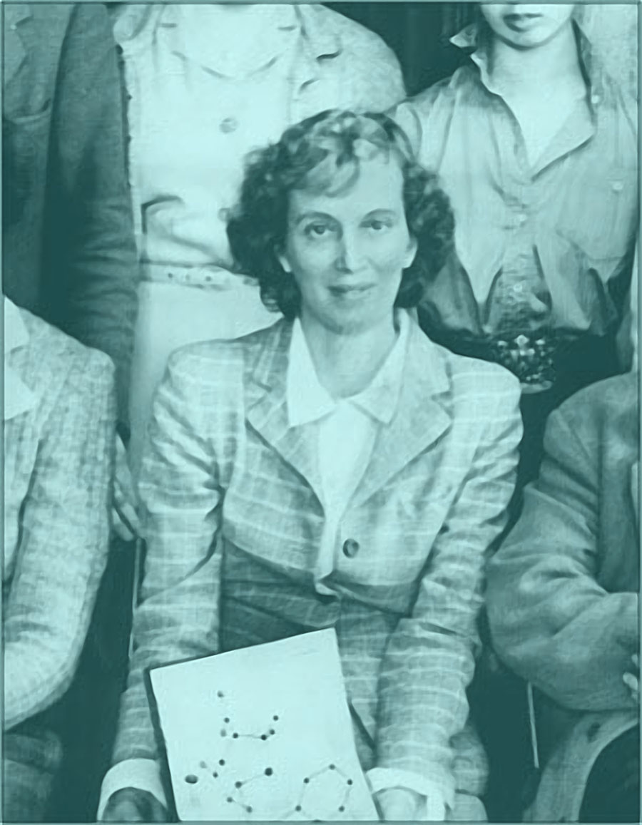
Nobel Prize winning British chemist Dr. Dorothy Hodgkin significantly contributed to unraveling the secrets of diabetes mellitus. She is considered a founder of X-ray crystallography, a technique used to discern the structures of organic biomolecules. Dorothy Hodgkin’s discovery of insulin's 3D structure paved the way for many scientific breakthroughs in diabetes research for years to follow. In support of International Women’s Day 2018, ALPCO is honoring the historic research of Dr. Dorothy Hodgkin.
 Dr. Dorothy Hodgkin’s childhood passion to understand mineral structures guided her to research and improve protein X-ray crystallography1,2. After many years of research, her hard work and dedication led her to discover the 3D structure of many important biomolecules, including porcine insulin3. Her cornerstone discovery of insulin's 3D structure has influenced further scientific breakthroughs, improved diabetes care, and inspired many scientists around the world.
About Dr. Dorothy Hodgkin
Dr. Dorothy (Crowfoot) Hodgkin (1910–1994) was born in Cairo, Egypt and spent most of her childhood in northern Africa and the Middle East1,2. She was highly curious about the different types of minerals in these regions1,2. Her parents were both British archeologists who supported her interest in the sciences1,2. Family friends also encouraged her fascination with the structures of minerals and crystals1,2. In 1937, Dr. Hodgkin’s passion for science drove her to earn a PhD from the University of Cambridge for her work on improving X-ray crystallography techniques and researching the chemistry of sterols3.Protein X-ray Crystallography
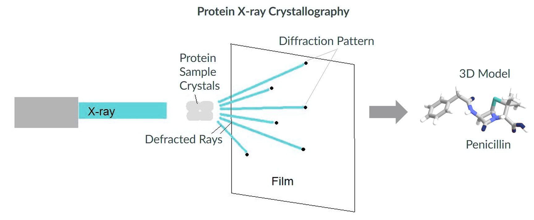
X-ray crystallography is a scientific technique that enables researchers to decipher the three-dimensional (3D) structures of biochemical compounds. X-ray beams pass though crystalized samples which generate specific diffraction patterns. These diffraction patterns are analyzed to determine the structure of the compound in the sample5. In protein crystallography, many factors can influence crystallization of the sample such as purity and concentration5. Therefore, the process of obtaining usable diffraction patterns of proteins can be very challenging5. Determining the structure of a protein, however, allows investigators to gain a better understanding of the protein and promotes further research into structure-based drug design, mechanism of action, and enzyme-substrate interactions5,6.
1964 Nobel Prize in Chemistry
As a researcher, Dr. Hodgkin used X-ray crystallography to determine the 3D structures of many important biochemical structures such as penicillin and vitamin B121,2. As a result, Dr. Hodgkin was awarded the 1964 Nobel Prize in Chemistry for her discoveries1,2 . Dr. Hodgkin was the third woman to ever receive the Nobel Prize in Chemistry, and remains the only British woman to have won this category9.Dorothy Hodgkin's Discovery of Insulin's 3D Structure
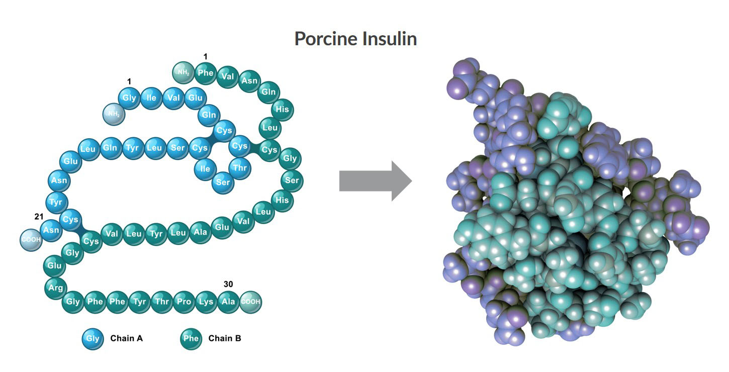
After 35 years of research, Dr. Hodgkin improved X-ray crystallography methods sufficiently to understand the complex 3D structure of porcine insulin9. Her research showed the overlay between the A Chain and B Chain in the porcine insulin molecule10. In this video below from the 1988 Nobel Laureates Symposium, Dr. Hodgkin lectures about her historical findings. https://youtu.be/pNl5rwIg4B4 The discovery of insulin’s 3D structure by Dr. Hodgkin led to more research breakthroughs such as insulin mutants and analogs11. The structures of insulin from other species were also identifiable with X-ray crystallography11. Ultimately, diabetes care has greatly improved as a result of determining porcine insulin’s 3D structure.
Dr. Dorothy Hodgkin’s childhood passion to understand mineral structures guided her to research and improve protein X-ray crystallography1,2. After many years of research, her hard work and dedication led her to discover the 3D structure of many important biomolecules, including porcine insulin3. Her cornerstone discovery of insulin's 3D structure has influenced further scientific breakthroughs, improved diabetes care, and inspired many scientists around the world.
References
- Science History Institute. (2017). Dorothy Crowfoot Hodgkin. sciencehistory.org.
- Nobel Lectures. (1972). Dorothy Crowfoot Hodgkin – Biographical, Chemistry 1963-1970. Elsevier Publishing Company, Amsterdam. nobelprize.org.
- Hodgkin. (1937). X -ray crystallography and the chemistry of the sterols. Cambridge University. Date: 27 April 1937. idiscover.lib.cam.ac.uk.
- Huydang2910. (2017). Hodgkin and penicillin structure. commons.wikimedia.org.
- Smyth & Martin. (2000). X-ray crystallography. Mol Pathol. 2000 Feb;53(1):8–14. PMCID: PMC1186895.
- Ennifar. (2013). X-ray crystallography as a tool for mechanism-of-action studies and drug discovery. Curr Pharm Biotechnol. 2013;14(5):537-50. PMID: 22429136.
- Kim. (2008). X-ray Crystallography. English Wikibooks. commons.wikimedia.org
- Cacycle. (2008). Chemical structure of Penicillin G. commons.wikimedia.org
- Houghton. (2014). Pioneer in X-ray crystallography, Dorothy Hodgkin is the only British woman to have received the Nobel Prize in chemistry. Royal Society of Chemistry. Rsc.org.
- Adams et al. (1969). Structure of Rhombohedral 2 Zinc Insulin Crystals. Nature. 01 November 1969;224:491–495. doi:10.1038/224491a0.
- Ward & Lawrence. (2011). Landmarks in Insulin Research. Front Endocrinol (Lausanne). 2011; 2:76. PMCID: PMC3356151.